Fig. 5.
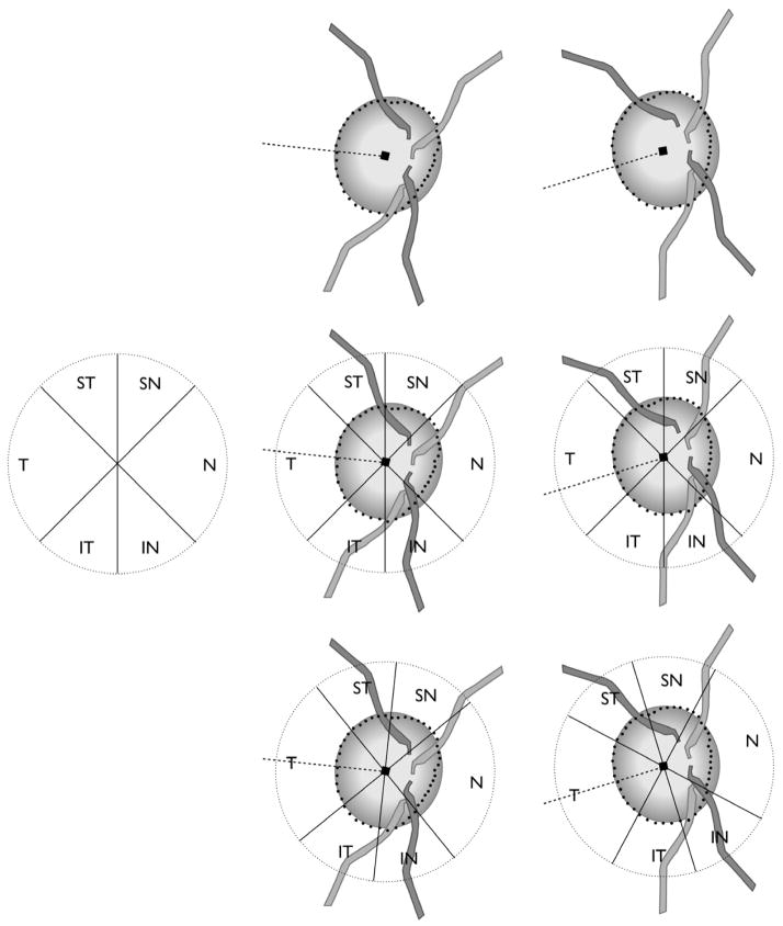
Impact of regionalisation of neuroretinal rim width and peripapillary retinal nerve fibre layer thickness measurements based on the axis connecting the fovea and Bruch’s membrane opening (BMO) centre (fovea-BMO centre axis). Top centre: eye with fovea-BMO centre axis of +6°. Top right: eye with fovea-BMO centre axis of −17°. Middle left: Sector regionalisation according to positions that are fixed relative to the imaging frame of the imaging device, currently applied to all eyes in the analyses of rim width and retinal nerve fibre layer thickness. Middle centre and right: Current regionalisation with fixed sector orientation leads to measurements from variably different anatomic locations. Bottom centre and right: Regionalisation relative to fovea-BMO centre axis. In this case rotating the sector orientation according to the fovea-BMO centre axis ensures that sectors contain measurements from the same anatomic locations.

