
Fig. 6.
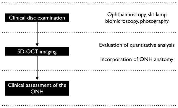
Schematic of incorporating spectral domain optical coherence tomography (SD-OCT) imaging that includes quantitative analyses and an understanding of optic nerve head (ONH) anatomy into a clinical examination of the optic disc with ophthalmoscopy, slit lamp biomicroscopy or photography to yield an clinically informed assessment of the ONH.