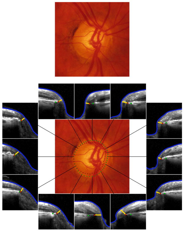
Fig. 8.
Incorporation of spectral domain optical coherence tomography (SD-OCT) into a clinical examination of the optic disc in a right eye suspected of glaucoma (top). In this case SD-OCT (bottom) with analysis with Bruch’s membrane opening minimum rim width (BMO-MRW) by clock hour provides valuable additional information. There is a considerable mismatch between the clinically visible disc margin (green dots) and BMO (red dots) indicating a clinically invisible extension of Bruch’s membrane, especially inferiorly and nasally. In these locations as well as the superior sector, which is the most suspicious, the neuroretinal rim is considerably thinner than the clinician would estimate from disc margin based evaluation. Future algorithms that indicate whether individual BMO-MRW values lie within or outside age-specific normal values could be clinically valuable.

