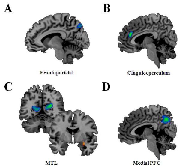
Figure 2.
Differences in spatial weighting of the networks between PTSD and control groups. Contrasts in red depict spatial weighting where PTSD is greater than controls, whereas contrasts in blue depict spatial weighting where PTSD is less than controls. Statistical maps are thresholded at p <. 05 corrected for multiple comparisons. Multiple comparison correction was relaxed the amygdala. (A) In the frontoparietal network there was decreased spatial weighting in the precuneus in PTSD versus controls. (B) In the cingulooperculum network there was decreased spatial weighting in the anterior cingulate in PTSD versus controls. (C) In the MTL (medial temporal lobe) network, compared to controls, the PTSD group had decreased spatial weighting in bilateral parieto-occipital and increased spatial weighting in the right amygdala. (D) In the Medial PFC (prefrontal cortex) network there was decreased spatial weighting in the posterior cingulate in PTSD compared to controls.

