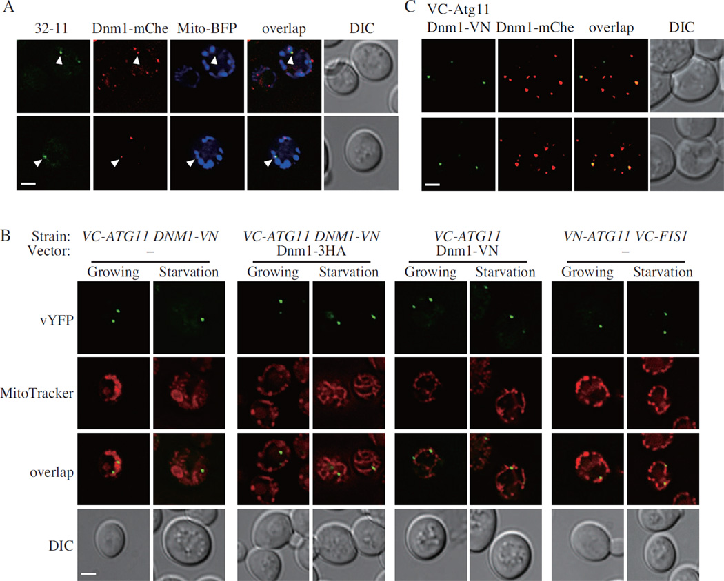
Figure 3. Atg11 Recruits Dnm1 to the Degrading Mitochondria.
(A) VN-ATG32 VC-ATG11 DNM1-mCherry cells, transformed with pMito-BFP, were cultured in SML and shifted to SD-N for 1 h, and samples were observed by fluorescence microscopy. Arrowheads indicate the colocalized 32-11 dots with Dnm1-mCherry on the mitochondrial reticulum. All of the images are representative pictures from single Z-sections. DIC, differential interference contrast. Scale bar, 2 µm.
(B) VC-ATG11 DNM1-VN cells transformed with empty vector or pDnm1-3HA, VC-ATG11 cells transformed with pDnm1-VN, and VN-ATG11 VC-FIS1 cells transformed with empty vector were cultured in SML and shifted to SD-N for 1 h. Samples were observed by fluorescence microscopy as in (A). Scale bar, 2 µm.
(C) VC-ATG11 DNM1-mCherry cells, transformed with pDnm1-VN, were cultured in SML and shifted to SD-N for 30 min. Samples were observed by fluorescence microscopy as in (A). Scale bar, 2 µm.
Also see Fig. S2.

