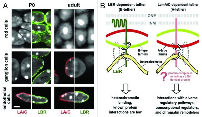
Figure 1. Heterochromatin clusters internally in the absence of peripheral tethers. (A) Temporal expression of LBR and lamin A/C in mouse retinal tissues. Tissues were stained simultaneously for lamin A/C (LA/C, red), LBR (green) and DNA (white, DAPI staining, where heterochromatin stains brightest). The distribution of one peripheral stain combined with DAPI is shown in each half-panel. Rod and ganglion cells express only LBR at stage P0; in adult tissue, ganglion cells have switched to lamin A/C (middle row) while rod cells uniquely express neither tether protein and exhibit the inverted nuclear architecture (top row). Endothelial cells express LBR and lamin A/C at both stages of development (bottom row). Scale bar = 2 µm. (B) Models for lamin A/C and LBR tethering of peripheral heterochromatin. Chromatin/DNA binding by lamins is indicated by dotted circles to show that lamins are not sufficient for heterochromatin tethering. From Solovei et al.4 with permission from Elsevier.

An official website of the United States government
Here's how you know
Official websites use .gov
A
.gov website belongs to an official
government organization in the United States.
Secure .gov websites use HTTPS
A lock (
) or https:// means you've safely
connected to the .gov website. Share sensitive
information only on official, secure websites.
