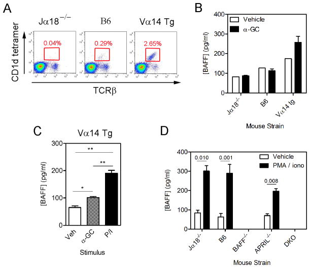
Figure 6. Expression of BAFF by bone marrow iNKT cells.
(A) Shows frequency of bone marrow iNKT cells in Jα18−/−, B6, and Vα14 Tg mice. (B–D) Bone marrow cells were cultured in triplicate as indicated before collection of supernatants and measurement of BAFF concentrations by ELISA. (B) Cells from Jα18−/−, B6, and Vα14 Tg mice were treated for 6 hr with vehicle or α-GC (C) Cells from Vα14 Tg mice were treated as in (B) except that PMA/ionomycin treatment was included. (B) and (C) are representative of three independent experiments that produced similar results. (D) Bone marrow cells from strains depicted were treated with vehicle or PMA/ionomycin. Data in B–D show mean ± SD BAFF concentration for triplicate samples. Data in (B) and (C) are representative of three independent experiments. (D) is from a single experiment using duplicate samples.

