Figure 1.
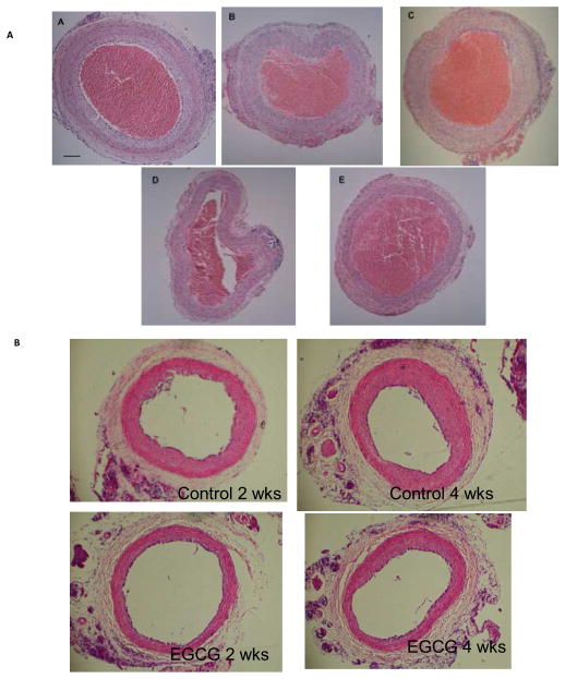
(A) Representative cross sections of rat carotid arteries harvested at two weeks post injury and stained with hematoxylin and eosin. Images show extent of intimal hyperplasia in the Control (A) and suppression by phytochemical inhibitors: Allicin (B), Resveratrol (C), SFN (D), and EGCG (E). Unflushed blood occupies the lumen of the specimens. Size bar represents 50 microns. n=5–6 for each group. (B) Representative cross sections of rat carotid arteries harvested at 2 or 4 weeks after injury, stained with H&E. n=6 for each group.

