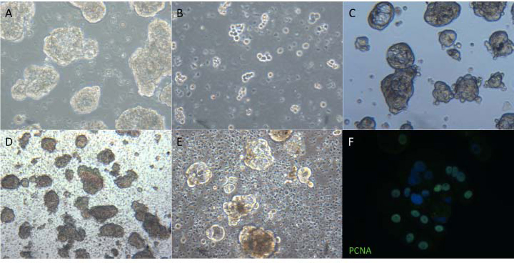
Fig. 3.
Subculture, cryopreservation, and proliferation of LuCaP cultured cells. Images of LuCaP 96 spheroids before (A), immediately following (B), and four days after digestion with trypsin/EDTA (C). Images of LuCaP 96 immediately following (D) and four days after thawing (E) as well as immunofluorescent detection of nuclear PCNA, indicating active proliferation (F). All images 100X.

