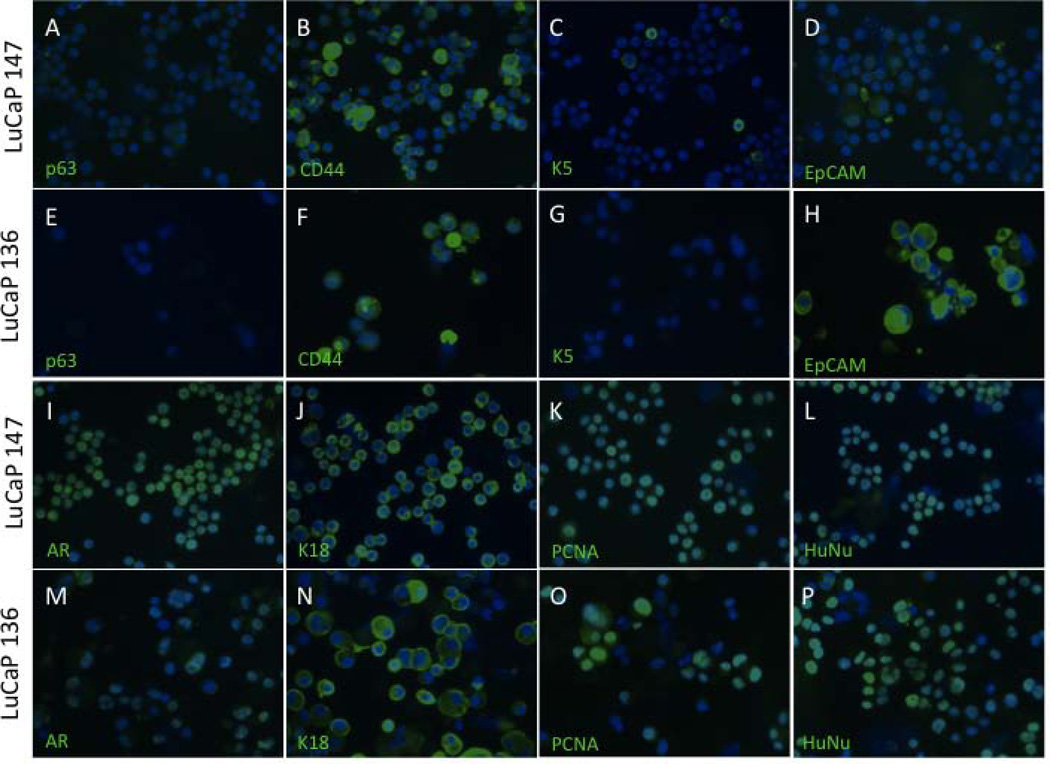
Fig. 5.
Immunofluorescence of prostate cell markers in cells from cultured LuCaP 136 and 147 spheroids. Cells lack expression of basal cell markers p63 (A and E) and K5 (C and G) but show heterogeneous staining for CD44 (B and F) and EpCAM (D and H). Cells are strongly positive for luminal cell markers AR (I and M) and K18 (J and N) as well as the proliferative marker PCNA (K and O) and HuNu (L and P), a marker of human nuclei.

