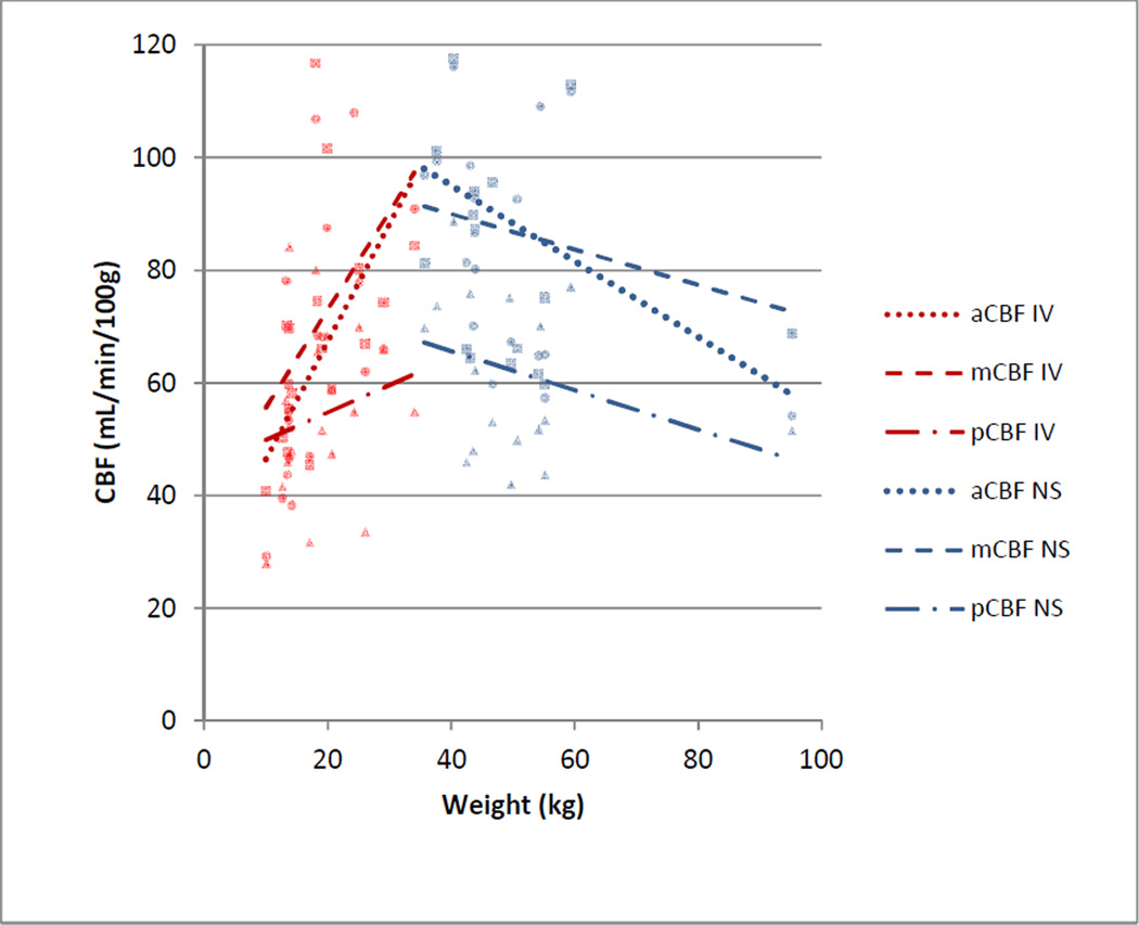
Fig. 4.
Cerebral blood flow (CBF) by weight in the anterior cerebral artery (aCBF, round data points), middle cerebral artery (mCBF, square data points), and posterior cerebral artery (pCBF, triangle data points) territories by group (IV= propofol, NS=non-sedated). CBF increased with weight in patients receiving propofol

