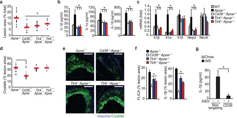
Figure 4. Inflammasome activity is impaired in atherogenic mice deficient in CD36 and its signaling partners TLR4 and TLR6.
Mice of the indicated genotype (Apoe–/–, Cd36–/–Apoe–/–, Tlr4–/– Apoe–/–, Tlr6–/–Apoe–/–, or control C57/BL6 Apoe+/+) were fed a western diet for 12 weeks, and atherosclerosis and inflammasome activity was assessed. (a) Lesion area in the aorta en face measured as a % of total aortic area. (b) Serum cytokine concentrations measured by Raybiotech Protein Array (IL1β, IL1α) or ELISA (IL18). (c) qRT-PCR analysis of aortic mRNA. (d-e) Plaque crystal content measured in serial sections throughout aortic root by combined confocal reflection microscopy illustrated in (e) and quantified in (d). (f) Plaque caspase-1 activity measured in serial sections throughout aortic root using FAM-YVAD-fmk FLICA probe fluorescence (left) or anti-IL-1β immunofluorescent staining (right) (n=8 sections/mouse) and expressed as % of total plaque area. (g) Serum IL-1β concentrations from Apoe–/– mice before (chow) or after western diet (WD) feeding for 4 wk during which mice were treated with a non-targeting or CD36 antisense oligonucleotide (ASO). Data is presented for n=15 mice (Apoe–/–), n=10 mice/group (Cd36–/–Apoe–/– and Tlr4–/–Apoe–/–) and n=7 mice (Tlr6–/–Apoe–/–) (a,d). Horizontal bars indicate the mean and symbols indicate individual mice. All other data is mean ± s.e.m. for n=4 mice/group (b-c), n=6 mice/group (f) and n=5 mice/group (g). Scale bar = 200 μm. *P<0.05.

