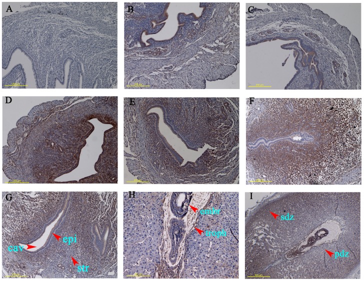
Figure 2. Immunohistochemical study showing the distribution of SPARC protein in mouse uteri during the early pregnancy.
Mouse uteri were collected on day 0 (B), 1 (C), 4 (D), 5 (E,F) and 8 (G,H) of pregnancy. A: negative control with normal serum was used in place of anti-SPARC antibody; E,G: inter-implantation sites; F,H: implantation sites; I is a shrunken picture of H. cav, uterine cavity; epi, endometrial epithelia; str, endometrial stroma; embr, embryo; troph, trophoblast; pdz: primary decidua zone; sdz: secondary decidua zone. Representative of three independent experiments.

