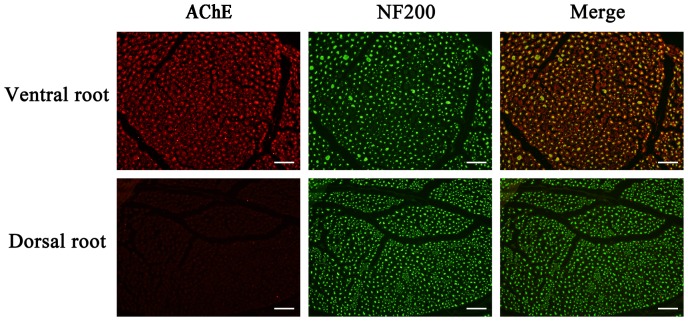
Figure 6. Differences in the axonal distribution of AChE between the ventral and dorsal roots.
The axons of the neurons are labeled with anti-NF200 (green). The AChE protein (red) was detected in the ventral roots, but was detected only at low levels in the dorsal roots. Merged images of AChE and NF200 staining are shown on the right. Scale bar, 50 µm.

