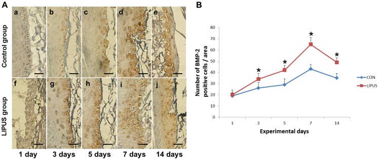
Figure 3. Quantitative analysis of BMP-2 expression.
(A). Effects of LIPUS stimulation on BMP-2 positive cells in the periodontium is shown by immunohistochemistry (bar: 20 μm). BMP-2 immunoreactivity was observed on the alveolar bone surface in the periodontium of the tension side in the LIPUS stimulation group (g–j) on days 3, 5, 7, and 14, significantly higher than that (b–e) in the non-stimulation group. (B). The number of BMP-2 positive cells in the LIPUS stimulation group was greater than that in the control group on days 3, 5, 7, and 14. *indicates P<0.05. Values are shown as the mean ± SD, and n = 4.

