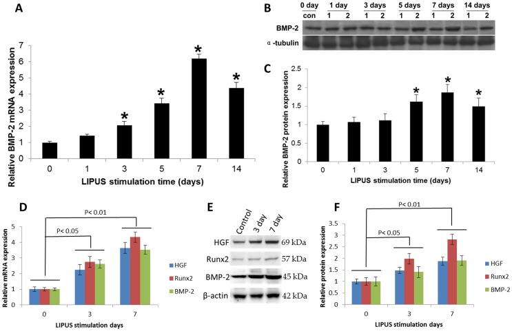
Figure 5. LIPUS stimulation enhanced the BMP-2 signaling pathway gene expression in vitro and in vivo.
(A). hPDL cells were cultured in the presence and absence of daily LIPUS stimulation. BMP-2 mRNA expression was determined using qRT-PCR. (B). BMP-2 protein (ROW 1; 45 kDa) can be detected in the control and LIPUS groups (1: CON; 2: LIPUS). The LIPUS groups showed higher expression of BMP-2 from day 5. (C). Quantification of BMP-2 protein expression in the LIPUS stimulation group was greater than that in the control group on days 5, 7, and 14. *indicates P<0.05. (D–F). Rat upper first molars were stimulated with or without LIPUS for different time intervals, and HGF, Runx2, BMP-2 were determined by qRT-PCR and Western blot, respectively. The data indicated that LIPUS increased HGF, Runx2, and BMP-2 mRNA (D: LIPUS stimulation 0 day vs LIPUS stimulation 3 day, P<0.05; 0 day vs 7 day, P<0.01) and protein expression (E, F: LIPUS stimulation 0 day vs LIPUS stimulation 3 day, P<0.05; 0 day vs 7 day, P<0.01) in vivo. The data are the mean ± SD of three separate experiments.

