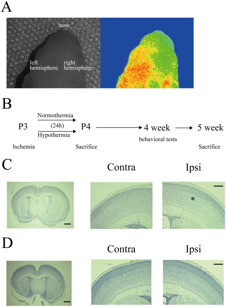
Figure 1. Examination of hypoxic ischemia injury in predevelopmental mouse brain.
A: Cerebral blood flow immediately after carotid artery ligation in a P2–P3 mouse. B: Experimental paradigm. Hypoxic injury was followed by 24 h of normothermia or hypothermia, and mice were sacrificed at the indicated time points (P4, 5 weeks). Note that behavioral tests were conducted at 4 weeks of age. C, D: Representative examples of hematoxylin staining for normothermia (C) or hypothermia (D) treated mice. In the normothermia group, the boundary was obscure between the superficial layers and the deep layers (white asterisk). Scale, 500 µm. High magnification images of the contralateral (left) and ipsilateral (right) hemisphere. Scale, 200 µm.

