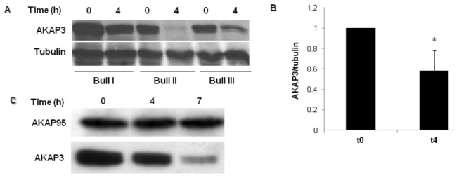
Figure 1. Detection of AKAP3 levels during capacitation.

(A) Bovine spermatozoa were incubated in capacitation medium. Samples were removed at the beginning and after 4h of capacitation. Proteins were extracted and analyzed by western blot using anti AKAP3 antibody and anti-α-tubulin antibody (loading control). (B) Densitometric analysis of AKAP3 bands. The graph represents an average of six different experiments ± SD at each time point. *Significance P< 0.01. (C) Bovine spermatozoa were incubated in capacitation medium. Samples were removed at the beginning, and after 4h and 7h of capacitation. Proteins were extracted and analyzed by western blot using anti-AKAP3 and anti-AKAP95 antibodies. The result represents three independent experiments.
