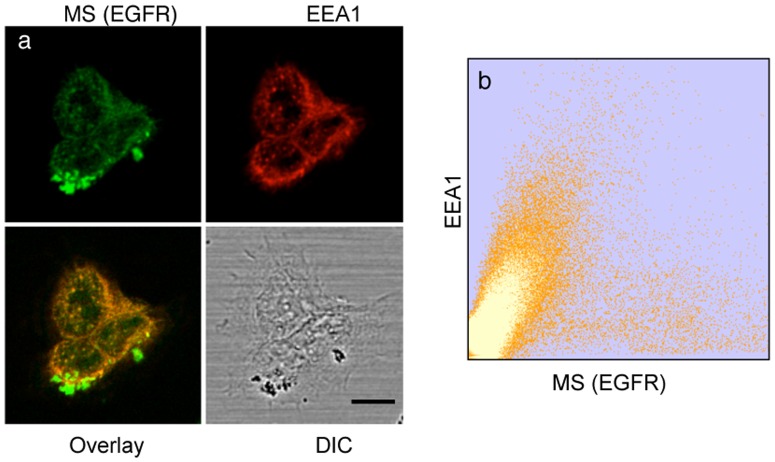
Figure 5. Localization of MS in endocytic vesicles after magnetic field pulse and 37°C incubation.
(a) Confocal microscopy images showing the endocytosis of MS after 3 min magnetization and subsequent incubation for 20 min at 37°C in the absence of magnetic field. Green, MS signal; red, immunofluorescence staining of early endosomes by Mab EEA1 and GAMIG-CY3. Scale bar is 10 µm. (b) Two-dimensional colocalization histogram of MS and EEA1 from a z stack (0.7 µm sections) of 20 images.

