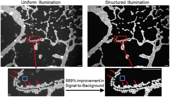
Figure 3. Images of a single layer of 10 µm fluorescent spheres embedded in PDMS and TiO2.
The reduced scattering coefficient of the phantom is approximately 10 cm−1. Images were taking using a 4×NA = 0.1 objective and an illumination frequency of 31.7 mm−1. The improvement in contrast is clearly seen from the uniform to structured illumination image. The signal-to-background was calculated by taking the intensity of 5 manually selected spheres (indicated by red arrows) and dividing by the background ROI (indicated by the blue square). All scale bars are 100 µm.

