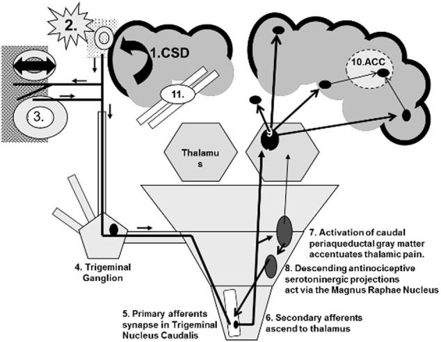
Figure 1.

Central sensitization using migraine as a model. (1) Cortical spreading depression (CSD) depolarizes cortical neurons and glia. (2) They release glutamate, K+, H+, metalloproteases, and other agents that dilate pial vessels and activate trigeminal nociceptive nerves. (3) The bifurcated neurons release calcitonin gene related peptide (CGRP) and other vasodilators near dural vessels by the axon response mechanism. Vascular wall stretching activates additional trigeminal nociceptive neurons (4) that have their primary synapse (5) in the upper cervical dorsal horn. (6) Ascending secondary afferents activate the thalamus. (7) Other afferents signal the periaqueductal gray matter. (8) Descending relays to the magnus raphae nucleus activate descending serotonergic neurons to inhibit the primary trigeminal (5 and 6) synapses. (9) Thalamocortical projections stimulate the hypothalamus, somatosensory cortex, amygdala, limbic system, and frontal cortex. (10) Pain, emotion, memory, frontal processing and other inputs converge on the anterior cingulate gyrus (ACC) and (11) interfere with its executive decision-making functions.
