
Table 2.
Regional Mean ADC Differences as a Function of Anatomic Location and Percentage VC
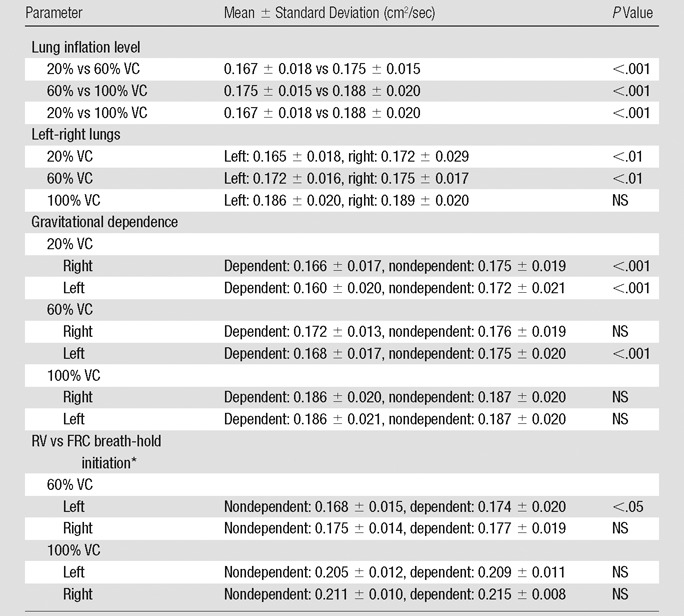
Note.—With respect to apical-basal lung regions, no differences in mean ADCs were observed at any lung volume. NS = not significant.
*
Data were obtained in the two subjects who underwent imaging at 60% and 100% VC following inspiration of 3He from FRC as opposed to RV.