
Figure 2.
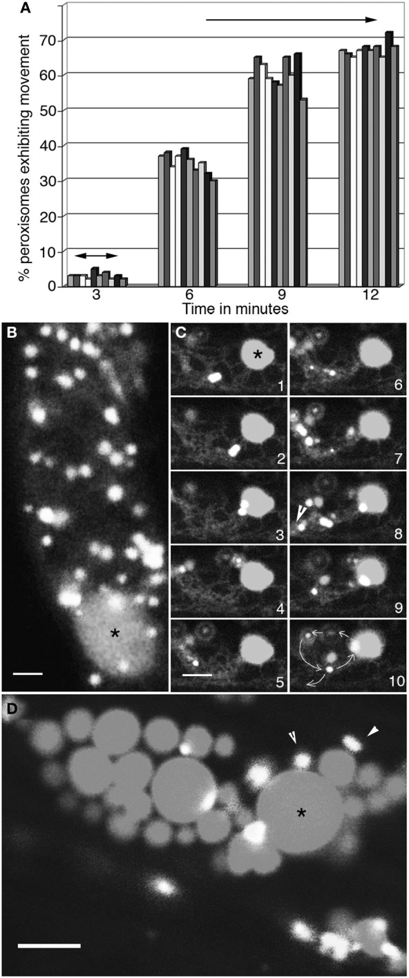
Concomitant changes take place in peroxisome and ER motility. (A) Graphical representation of changes in motility of 10 peroxisomes over 12 min as the ambient temperature around cells in 4°C treated seedlings rises to about 23°C. At 3 min following exposure to room temperature peroxisomes begin to exhibit short oscillations (double sided arrows) in synchrony with extension-retraction of ER tubules. The range of movement increases over time (6- and 9-min time points) until by 12 min both the ER and peroxisomes exhibit normal motility including long saltations. (B) Both peroxisomes and the ER in cells treated with 1μM latrunculin-B stop moving and large blobs of ER (*) surrounded by peroxisomes start appearing. (C) Sequential images taken at intervals of after washing away latrunculin-B show the gradual recovery of the ER accompanied by the circumambulation of peroxisomes apparently embedded in the ER (arrowhead in panel 8; path shown by circular arrows in panel 10) during the first 3–4 min. (D) Treatment with lat-B for more than 10 min usually leads to the formation of large ER globules (*) surrounded by static peroxisomes. Such disorganized ER does not reorganize easily into normal cytoplasmic streaming of organelles. Size bars = 5μm. *indicates an ER globule; arrowheads point to peroxisomes.