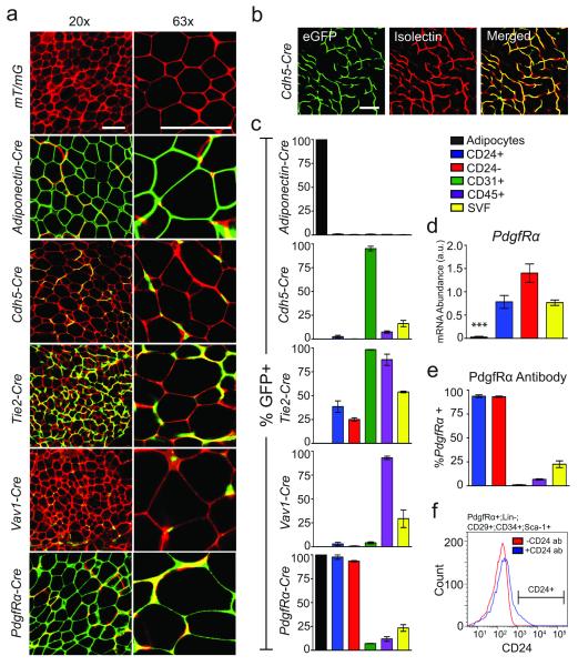
Figure 1.
Adipocytes are derived from PdgfRα+ precursor cells in subcutaneous WAT. (a) Confocal images of whole-mounted SWAT from indicated 4-week old Cre:mT/mG male mice (red: membrane-targeted dTomato; green: membrane-targeted eGFP, indicating Cre excision of dTomato). (b) Confocal images of membrane targeted eGFP and Isolectin GS-IB4 Alexa Fluor 647 staining endothelial cells of Cdh5-Cre:mT/mG SWAT. (c) Quantification of flow cytometry analysis of SVF populations from indicated 4-week old Cre:mT/mG mice (n=3). (d) Quantification of qPCR analysis of PdgfRα in mature adipocytes and FACS sorted SVF, Lin−:CD29+:CD34+:Sca-1+:CD24+ (CD24+) and Lin−:CD29+:CD34+:Sca-1+:CD24− (CD24−) cell populations (n=5 RNA extractions from independently isolated cell samples, *** p<0.001). (e) Quantification of flow cytometry analysis of anti-PdgfRα-PE antibody staining in indicated cell populations from 6-week old male C57BL/6 SWAT (n=3 SWAT SVF preparations). (f) A histogram of the distribution of CD24 staining in PdgfRα+:Lin−:CD29+:CD34+:Sca-1+ cells from (e). All error bars represent S.E.M. All scale bars represent 100 μm.

