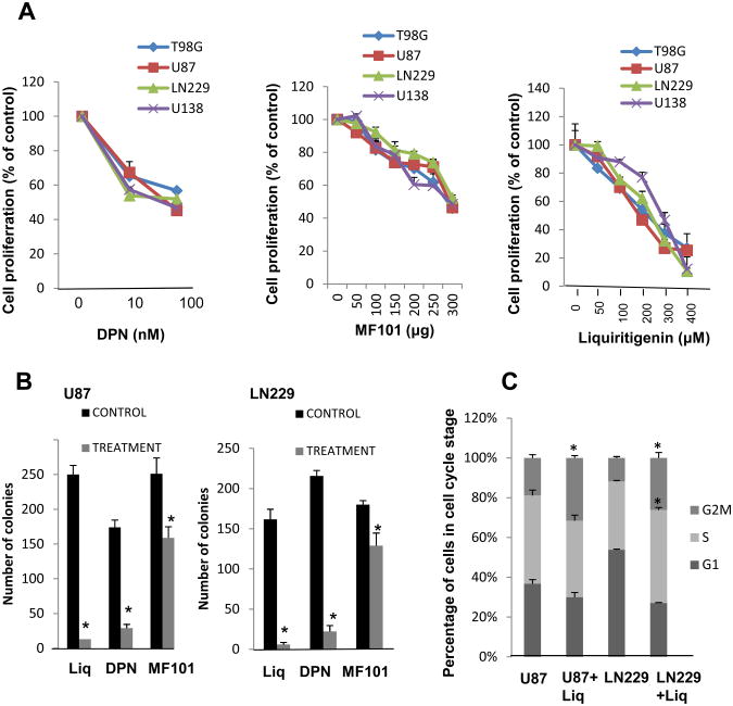
Fig. 3.
ERβ agonists inhibit the proliferation of glioma cell lines. A, T98G, U87, LN229, and U138 glioma model cells were treated with vehicle (0.1% DMSO) or indicated concentrations of DPN, MF101 and liquiritigenin for 72 h, and proliferation was measured using Cell Titer-Glo Luminescent Cell Viability Assay. B, U87 and LN229 cells were seeded in 6-well plates, and after 24 h the cells were treated with vehicle (0.1% DMSO) or DPN (1 μM), MF101 (250μg) and liquiritigenin (200 μM) for 72 h. After 7 days colonies were stained with crystal violet and colonies that contain ≥50 cells were counted. All data presented are the mean ± SEM. *, p< 0.05, t test. C, U87 and LN229 cells were treated with or without liquiritigenin (200 μM) and were subjected to flow cytometry. The percentage of cells in each cell cycle phase is shown in tabular form. All data presented are the mean of three experiments ± SEM. *, p< 0.05, t test.

