
Figure 4.
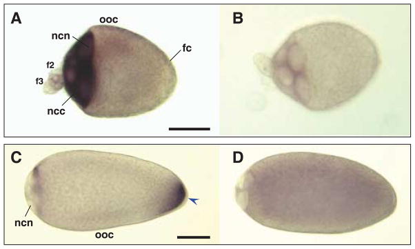
Hybridizations in situ of Culex quinquefasciatus nos and osk antisense and sense RNA probes to whole-mount oocytes and nurse cells. All primary follicles are orientated with the anterior on the left of the figure. Primary oocyte (ooc), with attached secondary (f2) and tertiary follicles (f3) dissected from ovaries of bloodfed Cx. quinquefasciatus females. Follicle cells (fc) surround the primary oocyte. Samples are hybridized with Cxqu nos antisense (A) and sense RNA (B) probes, or Cxqu osk antisense (C) and sense (D) RNA probes. (A) Strong staining is evident in the nurse cell cytoplasm (ncc) at stage IIIa and large nurse cell nuclei (ncn) are recognizable. (C) In stage IVa, oocytes appear elongated with a larger, bulb-shaped anterior end and the nurse cell nuclei occupy ~10% of the follicle length. Hybridization with Cxqu osk antisense RNA probes result in strong staining at the posterior end (blue arrowhead). (B, D) Stage IIIa and IVa oocytes hybridized with sense probes show no specific staining. Bar = 50 μM.