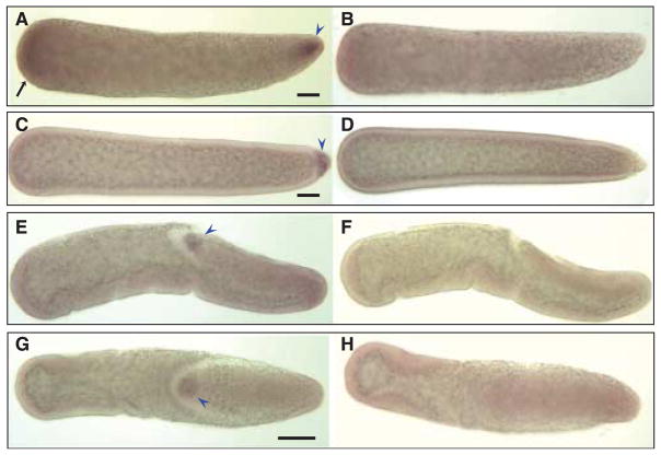
Figure 5.
Hybridizations in situ of Cxqu nos RNA probes to whole-mount Culex quinquefasciatus embryos. All embryos are orientated with anterior on the left of the figure. (A) A preblastoderm embryo hybridized with an antisense Cxqu nos RNA probe shows probe-specific staining at the anterior (black arrow) and posterior end (blue arrowhead), compared to a embryo hybridized with sense RNA probe (B). (C) Anterior staining appears to be transient and disappears after cellularization of the embryo; at this stage pole cells are stained strongly (blue arrowhead) when compared to a sense-probed embryo (D). Gastrulation stage embryos show specific staining of pole cells (blue arrowhead) in both lateral (E) and dorsal (G) views. F and H are the corresponding control embryos hybridized with sense-strand probes. Bar = 50 μM.

