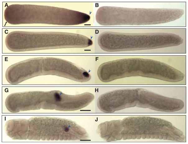
Figure 6.
Hybridizations in situ of Cxqu osk RNA probes to whole-mount Culex quinquefasciatus embryos. All embryos are orientated as in Figure 3. (A) A preblastoderm embryo hybridized with an antisense Cxqu osk RNA probe shows strong staining at the posterior end (blue arrowhead) and faint anterior (black arrow) staining when compared to a sense-probed embryo (B). (C) Transient anterior staining diminishes at blastoderm cellularization, and pole cells are stained strongly at this stage (blue arrowhead). (E) At the onset of gastrulation, pole cells are observed to lie on a cell plate and migrate dorsally (G) as germband elongation occurs. (I) In the final stages of embryonic development, pole cells have coalesced to the future gonad (blue arrowhead). Embryos hybridized with Cxqu osk sense probes exhibit no specific staining patterns (B, D, F, H, and J). Bar = 50 μM.

