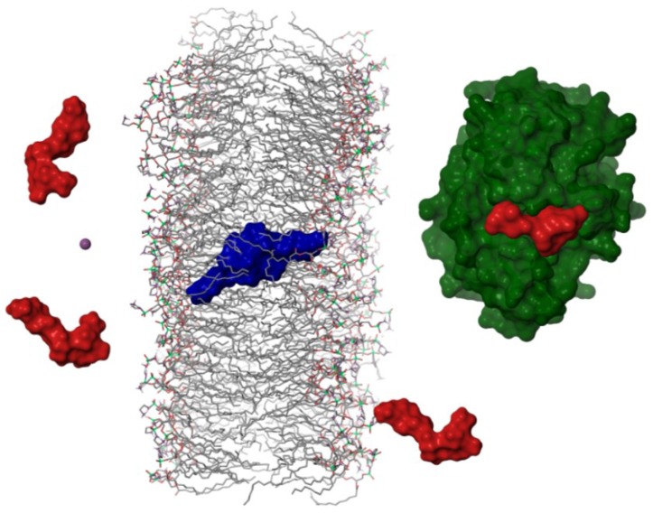
Figure 4.
Schematic representation of the hypothetical mechanism followed by okadaic acid to cross cellular membranes. Okadaic acid monomers (in red) would bind a potassium ion (purple sphere) from the extracellular media in order to form a dimer with a hydrophobic surface (in blue), that would cross lipid bilayers allowing the toxin to bind the catalytic subunit of protein phosphatases (PP2A in green [15]).

