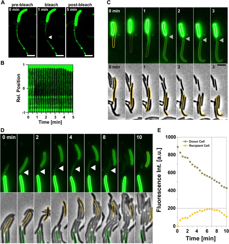
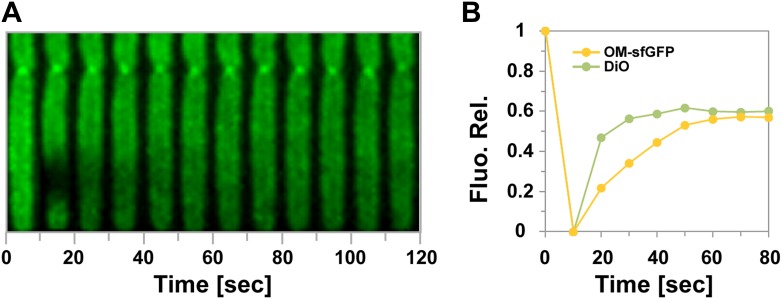
Figure 4. Transfer is driven by transient OM fusion between donor and recipient cells.
(A and B) Fluorescence recovery after photobleaching (FRAP) experiments targeting a tube connected to a single DiO+ cell. Rapid DiO exchange is observed between the tube and the cell body. The cell body is positioned at +1 in (A). (C) DiO transfer and formation of DiO+ tubes between two cells. An unstained recipient cell (orange cell contour) becomes stained in contact with a DiO donor cell (white cell contour). The grey arrow points to a tube formed between the two cells. Note that transfer only occurs between the two cells although other cells are also in contact with the donor cell. Scale bar = 1 µm. (D) DiO is exchanged by tubes connecting two cells. Fluorescence and corresponding phase contrast images between two transferring cells (green and orange contours) are shown. Scale bar = 1 µm. (E) Mean DiO fluorescence intensity over time in the donor cell (gray square) and the recipient cell (orange circle). The vertical dashed line represents the time where the tube connection was ruptured. The horizontal dashed line represents the maximal value of fluorescence intensity observed in the recipient cell.


