. 2013 Jul 23;2:e00953. doi: 10.7554/eLife.00953

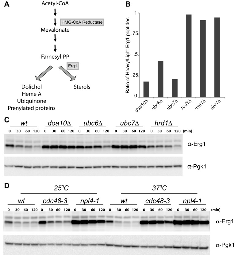
Figure 1. Erg1 is a substrate of the Doa10 complex.

(A) Schematic representation of the mevalonate pathway and its different end products. The steps catalyzed by HMG-CoA reductase and the squalene monooxygenase Erg1 are indicated. Adapted from Goldstein and Brown (1990). (B) Erg1 abundance in the indicated mutants relative to wt cells, as detected by mass spectrometry upon SILAC labeling. All strains used are lysine auxotrophs and were grown in the presence of either heavy L-lysine (wt cells) or light L-lysine (deletion mutants). Note that high steady state levels of Erg1 result in a low heavy/light ratio. (C) The degradation of endogenous Erg1 was followed after inhibition of protein synthesis by cycloheximide in wt cells or in cells with the indicated deletions. Whole-cell extracts were analyzed by SDS–PAGE and western blotting. Erg1 was detected with α-Erg1 antibody. Phosphoglycerate kinase (Pgk1) was used as loading control and detected with α-Pgk1 antibodies. A representative gel of three independent experiments is shown. (D) The degradation of endogenous Erg1 was analyzed as in (C) in wt cells or the temperature sensitive cdc48-3 and npl4-1 cells either at the permissive temperature of 25°C or after a 2 hr shift to 37°C, the restrictive temperature.

DOI: http://dx.doi.org/10.7554/eLife.00953.003

Figure 1.

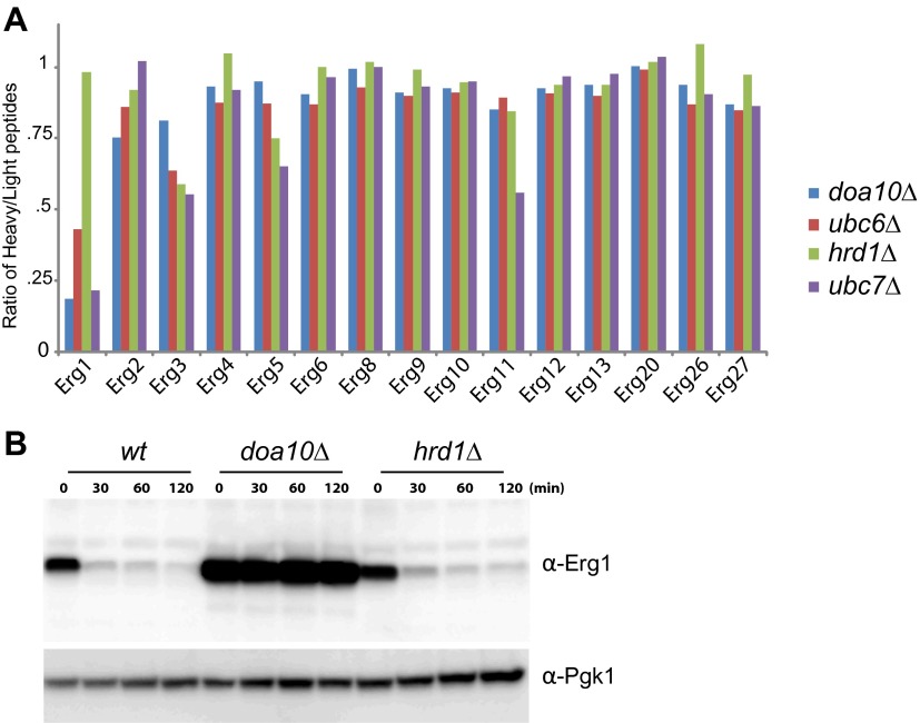
Figure 1—figure supplement 1. Abundance of Erg1 but not of other components of the Erg pathway is altered in Doa10 complex mutants.

Figure 1—figure supplement 1.

(A) Abundance of the indicated Erg pathway components in endoplasmic reticulum-associated protein degradation (ERAD) mutants relative to wt cells, as detected by mass spectrometry upon SILAC labeling. All strains used are lysine auxotrophs and were grown in the presence of either heavy L-lysine (wt cells) or light L-lysine (deletion mutants). Only proteins in which more than one peptide was detected by mass spectrometry were included. Note that high steady state levels of a protein result in a low heavy/light ratio. (B) The degradation of Erg1 was followed after inhibition of protein synthesis by cycloheximide in wt cells or in cells with the indicated deletions. Expression of Erg1 was driven from the strong constitutive promoter glyceraldehyde-3-phosphate dehydrogenase (GAPDH) replacing the endogenous ERG1 promoter. Whole-cell extracts were analyzed by SDS–PAGE and western blotting. Erg1 was detected with α-Erg1 antibody. Phosphoglycerate kinase (Pgk1) was used as loading control and detected with α-Pgk1 antibodies. A representative gel of three independent experiments is shown.