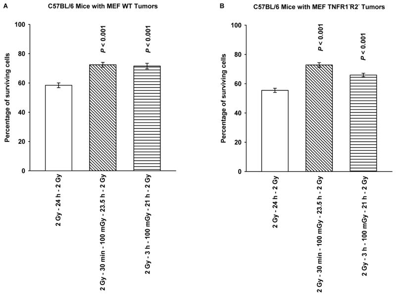
Figure 4.
Cell survival of MEF grown in the flanks of C57BL/6 mice as solid tumor growths, as a function of timing of a 100 mGy dose given 30 min or 3 h following the first of two 2 Gy doses separated by 24 h are presented for MEF WT (A) and MEF TNFR1−R2− (B). Both the first 2 Gy and subsequent 100 mGy doses were delivered to MEF growing in C57BL/6 mice. Tumors were removed just prior to the second 2 Gy dose, made into single cell suspension, and irradiated under in vitro conditions and surviving fraction assessed using an in vitro colony forming assay. P values were determined by comparing the survival of cells following two 2 Gy doses with those also exposed to a 100 mGy dose delivered 30 min or 3 h following the first 2 Gy dose using a Student’s two-tailed t test with values ≤ 0.05 identified as significant. Experiments were repeated 2x and error bars represent the SEM.

