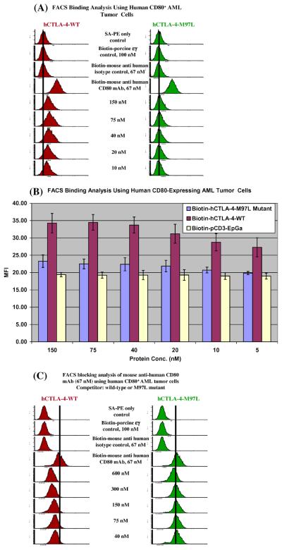
Fig. 1.
Human CTLA-4-M97L mutant almost lost the ability to bind to human CD80. (A) Flow cytometry binding analysis of human CTLA-4 wild-type (left panel) and human CTLA-4-M97L mutant (right panel) to the human CD80-expressing acute myelogenous leukemia cell line. Immunofluorescence staining was performed using biotinylated wild-type, mutant or anti-human CD80 mAb as the primary stain and PE-conjugated streptavidin (PE-SA) for the second stain. PE-SA only; biotinylated protein control [porcine CD3εγ ectodomain single-chain fusion protein (pCD3εγ)] [10]; isotype control [biotinylated mouse (BALB/c) IgG1, κ, clone# MOPC-21, Biolegend] were included. (B) Bar graph presentation of the mean fluorescence intensity (MFI) from flow cytometry binding analysis data as described in (A). Different concentrations of biotinylated pCD3εγ were used as non-specific fluorescence control. Error bars are included based on the calculated standard deviation. The hCTLA-4-M97L mutant's ability to bind to human CD80 was significantly decreased compared to the hCTLA-4-WT, p < 0.001. (C) Human CD80 blocking analysis by flow cytometry of the wild-type human CTLA-4 (left panel) and human CTLA-4-M97L mutant (right panel) with anti-human CD80 mAb (clone 2D10) to the human CD80-expressing acute myelogenous leukemia cell line. The concentration of the competitor is indicated. All figures of (A)–(C) are representatives of multiple individual experiments.

