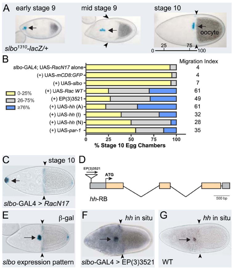
Fig. 1. Overexpression of hh rescues RacN17 border cell migration defects.

(A) slbo1310-lacZ/+ egg chambers at indicated stages stained for β-galactosidase activity (blue) to show border cells at beginning (left), during (middle) and end of migration when they reach the oocyte (right); migration distance indicated (bar). (B) Quantification of border cell migration at stage 10, with relative MI, following overexpression of indicated genes in a slbo-Gal4; UAS-RacN17 background (controls from (Geisbrecht and Montell, 2004)). Migration is shown as the percentage of border cells that migrated 0-25% (yellow), 26-75% (gray), or ≥76% (blue) of the distance to the oocyte. N > 100 egg chambers for each genotype. (C) Stage 10 slbo-Gal4; UAS-RacN17 egg chamber stained for slbo1310-lacZ; border cells did not migrate. (D) Diagram of the hh gene with coding exons (orange). EP(3)3521 is inserted 424 bp upstream of the ATG. (E) Stage 10 slbo1310/+ egg chamber stained to show slbo expression pattern (blue). (F, G) RNA in situ hybridization to detect hh expression at stage 10. (F) slbo-GAL4 driving EP(3)3521 shows increased hh expression in the slbo pattern. (G) Wild-type egg chamber. Arrows indicate border cells; arrowheads show extent of follicle cell rearrangement. Anterior to the left in this and subsequent figures.
