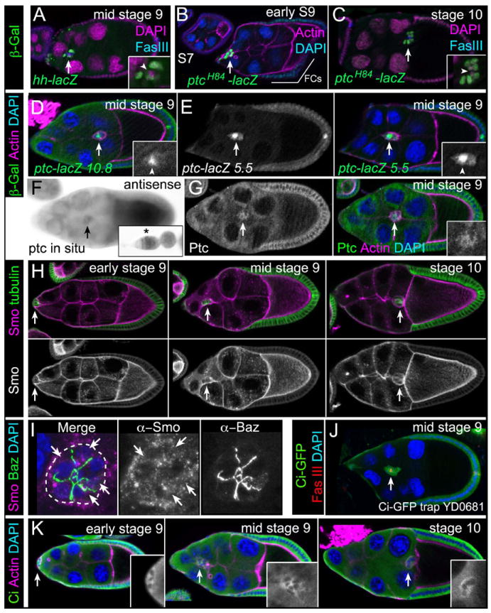
Fig. 2. Expression of Hh pathway components in wild-type border cells.

Representative images of egg chambers. Arrows indicate border cells. (A-E) Immunostaining for β-galactosidase (green) to detect hh-lacZ (A) or ptc-lacZ (B-E) enhancer trap (A-C) and promoter (D, E) reporters. Co-stains are Fas III (polar cells; blue in [A, C]), DAPI (nuclei; magenta in [A, C], blue in [B, D, E]) and F-actin (magenta [B, D, E]). Insets, magnified views of border cells (lacZ and DAPI in [A, C]); arrowheads, polar cells; FCs, follicle cells. (F) ptc mRNA expression. Inset, ptc expression in germarium. (G) Egg chamber immunostained for Ptc (green) and co-stained for F-actin (magenta). Inset, magnified view of border cells (Ptc). (H, I) Immunostaining for Smo (magenta). (H) Smo protein localization before, during, and after migration. α-Tubulin (green) labels all follicle cells. (I) Merged z-stack of border cells stained for Smo (magenta) and Bazooka (green). Smo localizes to cytoplasmic punctae (arrows); cluster outer border is outlined. (J, K) Ci protein (green) is present in border cells as detected by a GFP protein trap in ci (J) and using an antibody to Ci-FL protein (K; insets). Egg chambers were co-stained for Fas III (red [J]), F-actin (magenta [K]) and DAPI (blue).
