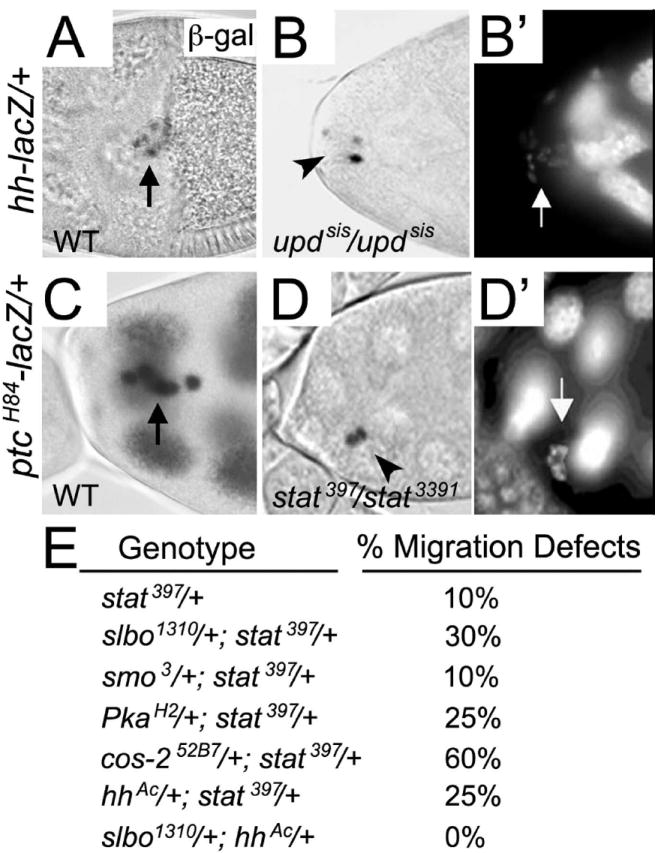
Fig. 3. JAK/STAT signaling regulates expression of hh and ptc in border cells.

(A-D’) Stage 10 egg chambers stained for nuclear lacZ activity in the border cells (arrows) and counterstained with DAPI (B’, D’) to mark nuclei. (A) LacZ reporter activity is present in all border cells in hh-lacZ egg chambers. (B, B’) Homozygous updsis mutant egg chamber in which the border cell cluster remained at the anterior tip of the egg chamber. Few border cell nuclei show hh-lacZ expression (arrowhead in B) even though border cells are present (arrow in B’). (C) LacZ reporter activity is present in all border cells in ptc-lacZ egg chambers. This egg chamber was slightly overexposed and thus staining in the nurse cells is more apparent. (D, D’) stat397/stat3391 mutant egg chamber in which border cells failed to migrate (arrowhead in D). ptc-lacZ expression is only present in two border cells, but all cells are visible by nuclear DAPI staining (arrow in D’). (E) Table showing results of dominant stat genetic interaction tests, expressed as the percentage of border cell migration defects found in trans-heterozygous flies of the indicated genotypes; 42 ≤ n ≤ 190 egg chambers for each genotype.
