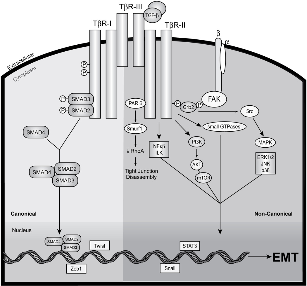
Figure 1.
Schematic depicting the canonical and noncanonical TGF-β signaling systems coupled to EMT in MECs. Transmembrane signaling by TGF-β ensues through its binding and activation of the Ser/Thr protein kinase receptors, TβR-I and TβR-II. Indeed, TGF-β binding to either TβR-III or TβR-II enables the recruitment and transphosphorylation of TβR-I, resulting its activation and subsequent phosphorylation the receptor-activated Smads, Smad2 and Smad3. Once activated, Smad2/3 form heterocomplexes with common the Smad, Smad4, which collectively translocate en masse to the nucleus to regulate the expression of TGF-β-responsive genes in concert with an ever expanding list of transcriptional coactivators and repressors. This branch of the bifurcated TGF-β signaling system represents the “canonical” or “Smad2/3-dependent” TGF-β pathway (left). Alternatively, TGF-β also activates a variety of “noncanonical” or “Smad2/3-independent” effectors, including Par6, NF-κB, ILK, FAK, Src, a variety of small GTPases, members of the MAP kinase family (e.g., ERK1/2, JNK, and p38 MAPK), and the PI3K:AKT:mTOR signaling axis (right). Activation of members of the Snail, ZEB, or bHLH family of transcription factors promote EMT by transcriptionally repressing the production of E-cadherin transcripts (bottom). Imbalances between canonical and noncanonical TGF-β signaling pathways have recently been identified and established as casual aberrancies underlying the initiation of oncogenic TGF-β signaling and its initiation of EMT in normal and malignant MECs. See text for details.

