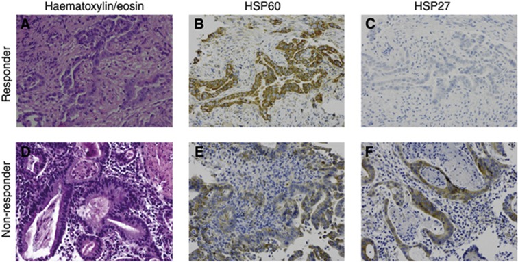
Figure 2.
IHC staining for HSP60 and HSP27 in pretherapeutic biopsies of two oesophageal adenocarcinoma cases. (A–C) a CTX responder (No.745, TRG 1) and (D–F) a non-responder (No.772, TRG 3), are shown ( × 200): (A) haematoxylin/eosin; (B) very strong HSP60 expression; (C) no HSP27 expression; (D) haematoxylin/eosin; (E) weak HSP60 expression; and (F) strong HSP27 expression.

