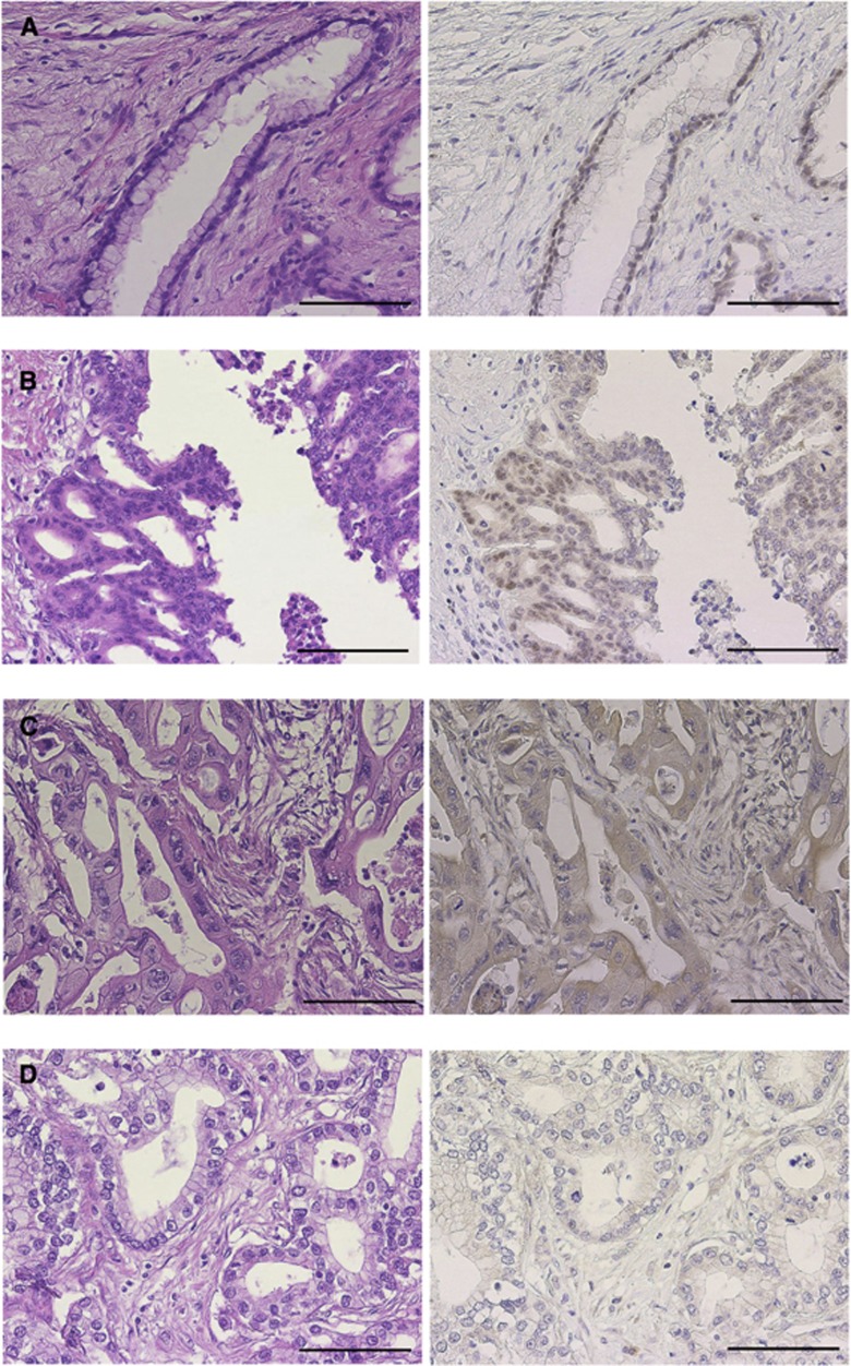
Figure 4.
Immunohistochemistry for SMARCC1 in clinical samples. (A–D) Haematoxylin and eosin staining on the left side and SMARCC1 staining on the right side. (A) A normal pancreatic duct sample. SMARCC1 expression was identified in the nucleus homogeneously in normal pancreatic duct cells. (B) A representative SMARCC1-positive sample. SMARCC1 staining was in the spotted granular nuclear pattern in pancreatic carcinoma cells. (C, D) Representative SMARCC1-negative samples. SMARCC1 staining was in the cytoplasmic pattern (not stained in the nucleus) or in the negative pattern (not stained in the nucleus and the cytoplasm) in pancreatic carcinoma cells. Bar=100 μm.

