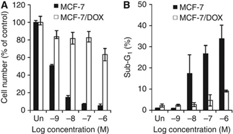
Figure 1.
The effect of paclitaxel on MCF-7/DOX cell survival. (A) MCF-7 and MCF-7/DOX cells were treated with increasing concentrations of paclitaxel ranging from 1 nℳ to 1 μℳ for 48 h, and the viable cell numbers were determined using trypan blue staining. (B) MCF-7 and MCF-7/DOX cells treated as in (A) were assayed for apoptotic population by flow cytometric analysis of the sub-G1 population. All quantitative data are shown as the means±s.d. from three independent experiments.

