Figure 5.
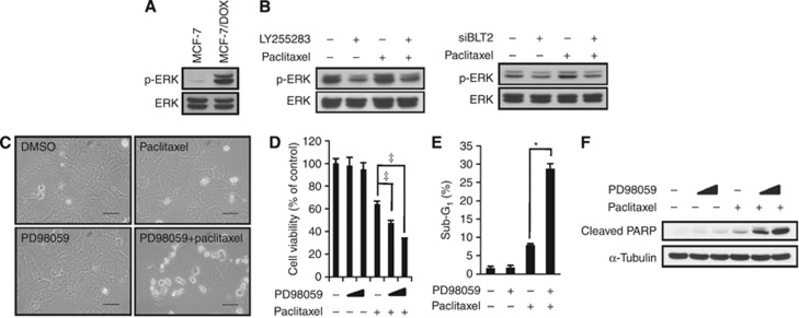
ERKs are downstream of BLT2 and mediate paclitaxel resistance in MCF-7/DOX cells. (A) Immunoblot analyses were performed to detect ERK phosphorylation in MCF-7 and MCF-7/DOX cells. The data are representative of three independent experiments. (B) MCF-7/DOX cells were pre-treated with LY255283 (10 μℳ) for 30 min before paclitaxel (1 μℳ) treatment for 6 h (left panel), or the cells were transfected with BLT2 or control siRNAs for 24 h before paclitaxel (1 μℳ) treatment for 6 h (right panel). The cells were then subjected to immunoblot analysis as in (A). The data are representative of three independent experiments. (C) MCF-7/DOX cells were pre-treated with PD98059 (10 μℳ) for 30 min before paclitaxel (1 μℳ) treatment for 48 h. Cell morphology was visualised using an Olympus BX51 microscope at × 40 magnification. Scale bars, 50 μm. (D) MCF-7/DOX cells were pre-treated with PD98059 (10 or 20 μℳ) for 30 min before paclitaxel (1 μℳ) treatment for 48 h. Viable cells were detected using an MTT assay. (E) MCF-7/DOX cells were pre-treated with PD98059 (10 μℳ) for 30 min and were then treated with or without paclitaxel (1 μℳ) for 48 h. Apoptotic cells were quantified by flow cytometric analysis of the sub-G1 population. All quantitative data are the means±s.d. of three independent experiments. *P<0.05, ‡P<0.005. (F) MCF-7/DOX cells treated as in (D) were assayed for apoptosis levels, as determined by PARP cleavage. The data are representative of three independent experiments.

