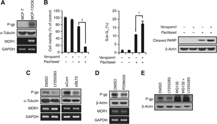
Figure 6.
The ‘BLT2–ERK' cascade regulates MDR1 protein levels in MCF-7/DOX cells. (A) Immunoblot assays and semi-quantitative RT-PCR analyses of P-gp and MDR1 mRNA were performed using MCF-7 and MCF-7/DOX cells. (B) MCF-7/DOX cells were pre-treated with verapamil (10 μℳ) for 30 min and were then treated with or without paclitaxel (1 μℳ) for 48 h. Cell viability and the percentages of cells in the sub-G1 population were determined using an MTT assay (left panel) and flow cytometric analysis (centre panel). In addition, cleaved PARP was determined using an immunoblot assay (right panel). All quantitative data are the means±s.d. of three independent experiments. †P<0.01, ‡P<0.005. (C) MCF-7/DOX cells were incubated with LY255283 (10 μℳ) or DMSO for 12 h, or transfected with BLT2 or control siRNAs for 48 h, and then subjected to immunoblot analysis and semi-quantitative RT-PCR of P-gp and MDR1 mRNA. (D) MCF-7/DOX cells were treated with PD98059 (10 μℳ) for 8 h, after which the amounts of P-gp and MDR1 mRNA were determined using an immunoblot assay and semi-quantitative RT-PCR. (E) MCF-7/DOX cells were incubated in the presence of 0.5% serum for 3 h and then incubated with LY255283 (10 μℳ) plus MG132 (10 μℳ) for 12 h. Cell lysates were subjected to immunoblot analysis with antibodies against P-gp and β-actin (loading control).

