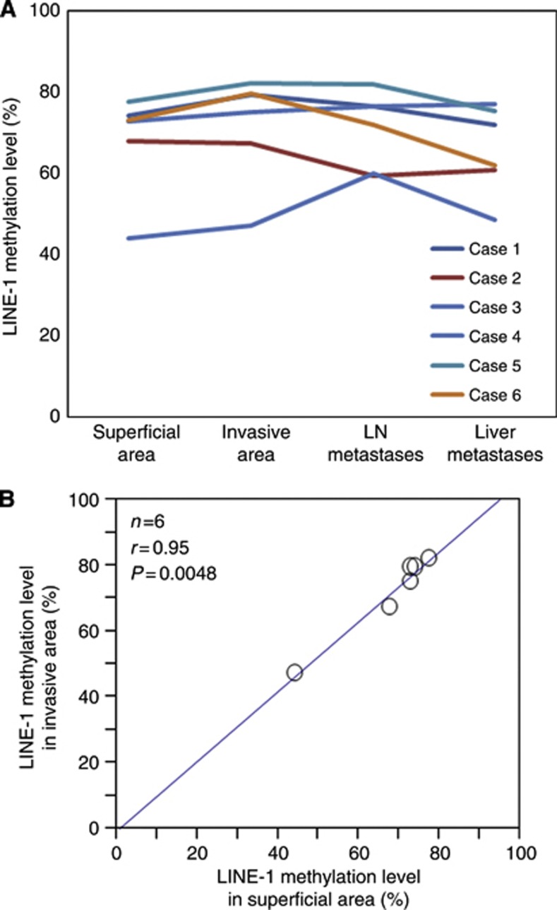
Figure 3.
LINE-1 methylation level in superficial and invasive areas of primary tumours, matched LN metastases, and matched liver metastases (n=6). (A) In each case, the LINE-1 methylation level of the LN and liver metastases was similar to that in the matched primary tumours. (B)The LINE-1 methylation level in superficial areas was significantly associated with that in the matched invasive areas within primary tumours (n=6; Pearson's correlation coefficient, r=0.95; P=0.0048).

