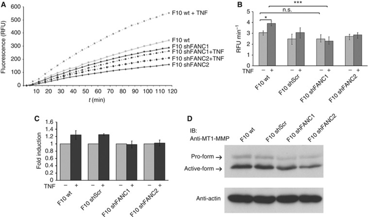
Figure 7.
The TNF-induced metalloproteinase activation is mediated by FAN. (A) One representative assay of MMP activation. Active MMPs cleave OmniMMP RED fluorogenic substrate (Enzo) emitting a fluorescent signal. The B16 F10 cells were incubated with the substrate. Increasing fluorescence is measured in RFUs over the course of 2 h. (B) The increase of fluorescence was determined as RFU min−1 and used as quantification of enzyme activity. (C) To visualise the induction, the stimulated TNF activity (RFU min−1) was normalised to the unstimulated value. Bars show mean values of three individual experiments, each performed in triplicate. Results are expressed as mean±s.e.m. (D) Cell lysates from B16 F10 melanoma cells were prepared, separated by SDS–PAGE and analysed by western blotting using an anti-MT1-MMP antibody. Actin probing ensured equal loading. Statistical analyses were standard two-tailed Student's t-test for two data sets using Prism (GraphPad Inc.). The P-values of <0.05 (*), <0.01 (**) and <0.001 (***) were deemed as significant, highly significant, and most significant, respectively.

