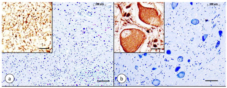
Figure 2. Dorsal nucleus of Clarke’s column in Friedreich’s ataxia.

(a) Friedreich’s ataxia; (b) normal control. (a) and (b) Cresyl Violet; insets: immunohistochemistry of class-III-β-tubulin. The normal dorsal nucleus displays large round nerve cells with peripherally located chromatin (b). The nerve cells have a few coarse dendrites (inset in [b]). In Friedreich’s ataxia (a), large neurons are absent. Class-III-β-tubulin reaction product is present in only a few neuritic processes that may represent the remaining afferent fibers (a, inset). Bars: (a) and (b), 100 μm; insets, 20 μm.
