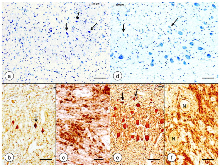
Figure 6. The dentate nucleus in Friedreich’s ataxia.

(a)-(c) Friedreich’s ataxia; (d)-(f) normal control. (a) and (d) Cresyl Violet; (b) and (e) immunohistochemistry of neuron-specific enolase; (c) and (f) immunohistochemistry of synaptophysin. In Friedreich’s ataxia, the large neurons of the dentate nucleus have disappeared (a). The small nerve cells (a, arrows) do not represent atrophic large neurons. Small neurons are also present in the normal dentate nucleus (d, arrows). They display strong reaction product with anti-neuron specific enolase (b and e, arrows). The synaptophysin stain in Friedreich’s ataxia (c) shows coarse clusters of modified corticonuclear terminals termed grumose degeneration. In the normal dentate nucleus (f), synaptophysin reaction product labels much smaller axosomatic and axodendritic terminals and generates negative images of normal neurons (N). Bars: (a-b), (d-e), 100 μm; (c) and (f), 20 μm.
