
Figure 4.
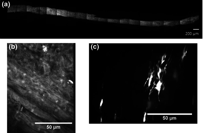
Characterization of fatigue damage in the tendon fascicle (bovine extensor tendon fascicle after 300 cycles of creep loading) using a combination of high-concentration acridine orange staining for collagen structure and calcein AM for cellular morphology: (a) low-magnification composite image of the fascicle length, (b) matrix structure after 300 cycles of creep loading and (c) calcein AM staining of tenocytes within the tendon fascicle after fatigue loading.