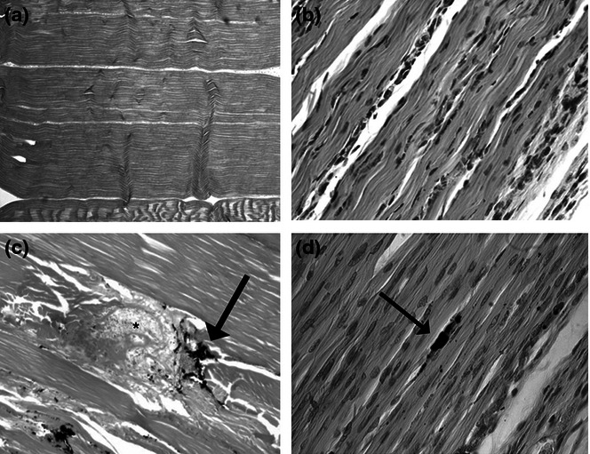
Figure 1.

H&E stain micrographs of normal adult tendon (a, 50× magnification) and normal fetal tendon (b, 400× magnification). Injured adult tendon (c) retains disruption of the collagen fibers at the site of India ink marking (arrow) and the disruption of normal collagen organization and disorganized granulation tissue at the site of wounding (asterisk). Injured fetal tendon (d) remodels, revealing no structural abnormalities or lack of collagen fiber disorganization at the wound site (identified with charcoal, arrow). The collagen architecture appears to be completely restored (adapted from Beredjiklian et al. 2003).
