
Figure 3. Spreading of hα-syn is associated with neuritic pathology.
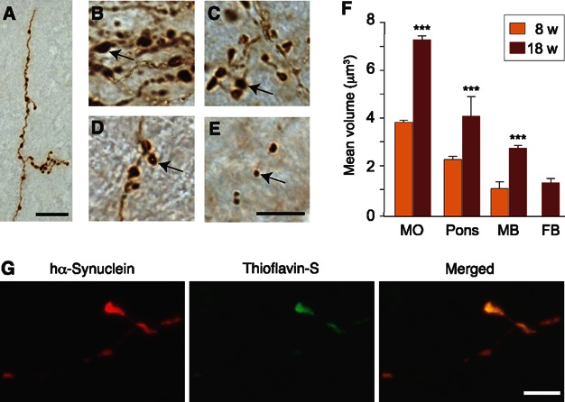
- A.Brain sections from a high expressor rat killed 18 weeks after vagal injection were stained with an anti-hα-syn antibody. The representative image shows a pontine axon with intensely stained swellings. Scale bar, 20 μm.
- B–E.Hα-syn-immunoreactive neuritic varicosities in different brain regions at 18 weeks post injection. Arrows indicate swellings of different sizes in the MO (B, 7.3 μm3), pons (C, 4.1 μm3), midbrain (D, 2.8 μm3) and forebrain (E, 1.4 μm3). Scale bar, 10 μm.
- F.The volume of hα-syn-immunoreactive neuritic swellings was measured in the MO (n = 1568 and 2406 swellings at 8 and 18 weeks, respectively), pons (n = 357 and 461 at 8 and 18 weeks), midbrain (n = 8 and 296 at 8 and 18 weeks) and forebrain (n = 34 at 18 weeks). Mean ± SEM. ***P < 0.001 by Wilcoxon Rank Sums test.
- G.Confocal images of a pontine axon stained with an anti-hα-syn antibody (red) and Thioflavin-S (green). Merged images show co-localization. Scale bar, 5 μm.