
Figure 1.
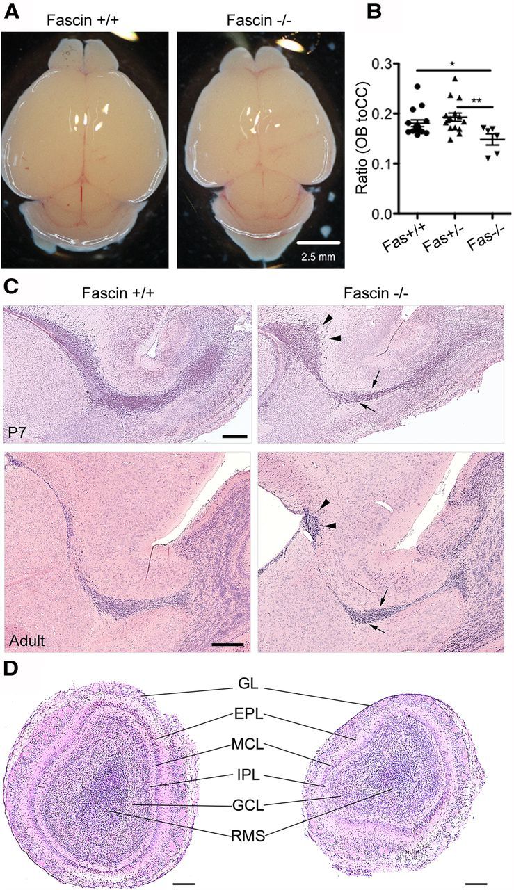
Fascin-1ko mice show a smaller OB and an abnormal RMS organization. A, B, Early postnatal (P7) homozygous fascin-1ko mice have a smaller brain (A) and a lower ratio between OB and cerebral cortex (CC) length (B) compared with wt and heterozygous littermates (mean ± SEM; n = 15 brains for wt and heterozygous fascin-1ko; n = 6 brains for homozygous fascin-1ko; *p < 0.05; **p < 0.01). C, Hematoxylin/eosin-stained sagittal brain slices from P7 (top row) and P50 (bottom row) mice showing abnormal RMS organization in fascin-1ko animals. Note the thinner RMS rostral section (arrows) and a caudal cell accumulation (arrowheads). D, Hematoxylin/eosin-stained coronal OB sections in P7 mice. The general organization of the OB appears preserved in fascin-1ko animals. GL, Glomerular layer; EPL, external plexiform layer; MCL, mitral cell layer; IPL, internal plexiform layer; GCL, granule cell layer. Scale bars: A, 2.5 mm; C, D, 200 μm.